Seeing mucosa with unprecedented cellular and molecular perspectives through endoscopy
Challenges - Esophageal Cancer
Esophageal Cancer
Each year, more than 600,000 new cases of esophageal cancer are diagnosed and more than 540,000 people die of this disease worldwide, making it the tenth most commonly diagnosed cancer and the sixth leading cause of death from cancer. Esophageal cancer is deadly with the second lowest survival rate of all cancers because the symptoms occur late in the course of disease. In the Western world, 50% of esophageal cancers are adenocarcinoma and its incidence has dramatically increased in the last 3-4 decades. Europe has the second highest incidence and mortality of esophageal cancer by volume compared with other continents. It is estimated that ~2% of all European citizens has Barrett’s esophagus, a well identified pre-cancerous stage of esophageal adenocarcinoma.
Finding esophageal cancer at an early stage
Endoscopic surveillance programs are implemented on Barrett’s esophagus patients to detect cancer early. Studies show that treatment of pre-symptomatic adenocarcinoma markedly improves patient outcome. Moreover, an identifiable transition phase between Barrett’s esophagus and adenocarcinoma known as dysplasia also offers a stage at which cancer can be prevented with endoscopic interventions. However, the current Barrett’s esophagus surveillance program is not effective at finding early lesions (dysplasia and cancer) due to the wide use of while light endoscopy, a technology insensitive to early lesions as they can be patchy , confined to very small areas and appear macroscopically indistinguishable from non-dysplastic mucosa.
As a result, quadratic biopsies are taken every 2 cm within the Barrett’s esophagus segment2 to maximize the chance of detecting early cancer. However, this method samples only a small fraction of Barrett’s esophagus, leading to miss 30% to 50% early lesions.
Vision
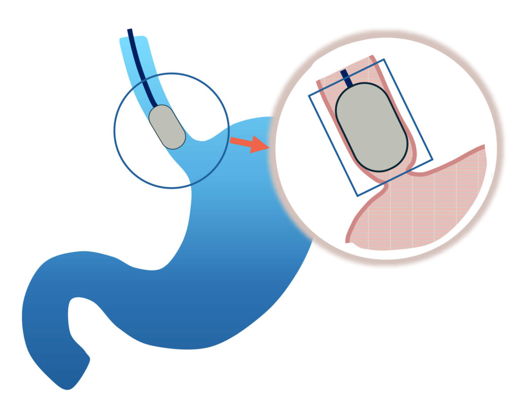
By developing several imaging techniques within the new endoscope, ESOHISTO aims to deliver revolutionary endoscopic technologies to highlight early lesions with high sensitivity through three-dimensional (3D) morphological and pathophysiological imaging, deciphering cell architecture, morphology and molecular information for diagnosis.
Optical coherence tomography (OCT)
Optical coherence tomography (OCT) is a high-resolution imaging technique with most of its applications in medicine and biology. OCT uses coherent light to obtain micrometer-level depth resolved images of biological tissue. It uses interferometric techniques to detect the amplitude and time-of-flight of reflected light to reconstruct 3D tissue images.
Optoacoustic imaging
Optoacoustic imaging (OI) is a novel multi-scale optical imaging method with a great potential in medicine and biology due to its unique ability of high-resolution imaging at depths beyond all other optical methods. To achieve this, OI uses modulated light to induce high-frequency optoacoustic waves generated from optical absorbers inside biological tissues. Applications include in vivo imaging of vascular morphology and oxygen saturation based on endogenous tissue absorbers, and molecular imaging based on various exogenous contrast agents.
Fluorescence microscopy
Fluorescence microscopy uses the ability of fluorochromes to emit light after being excited with light of a certain wavelength to construct high-resolution images. Using a confocal gating, 3D images of the expression of specific molecules marked by various fluorescent probes can be generated to facilitate early diagnosis.
Latest News:
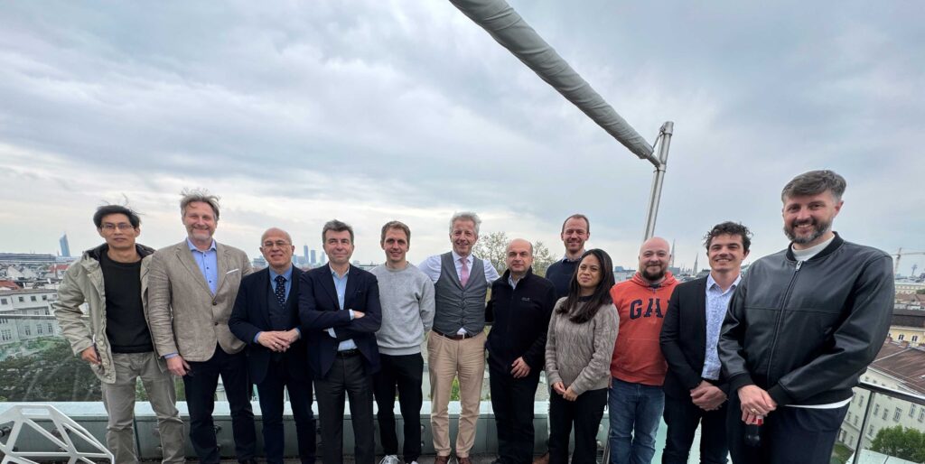
April 21st, 2026: Review meeting
The first review meeting of the ESOHISTO project was successfully hosted in Vienna on April 21, 2026.
Our project officer and expert reviewers visited the labs and engaged in thorough and thought-provoking discussions with the Consortium on the progress achieved in the first reporting period (M1 – M12). Thanks to their deep insights on helping shape ESOHISTO!
ESOHISTO is developing a groundbreaking hybrid endoscope designed to deliver real-time, 3D cellular and molecular imaging—paving the way for earlier and more precise detection of gastrointestinal cancers.
With a strong first year behind us, the Consortium is excited to move forward into the next phase of the project and continue to push the boundaries of in vivo histology and bring this transformative technology closer to clinical reality.